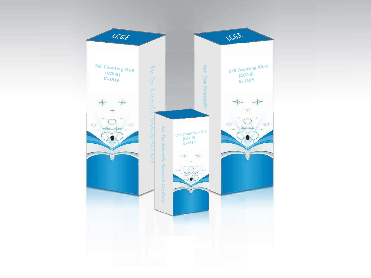
Cell Counting Kit-8 (CCK-8) IC-1519
BackGround
Cell Counting Kit-8 (CCK-8) provides a tool for studying induction and inhibition of cell proliferation in any in vitro model. Cell Counting Kit-8 (CCK-8) allows very convenient assays by utilizing highly water-soluble tetrazolium salt. WST-8 [2-(2-methoxy-4-nitrophenyl)-3-(4-nitrophenyl)-5-(2,4- disulfophenyl)-2H-tetrazolium, monosodium salt] produces a water-soluble formazan dye upon reduction in the presence of an electron mediator.
CCK-8 is a one-bottle solution, ready to use. CCK-8, being nonradioactive, allows sensitive colorimetric assays for the determination of the number of viable cells in cell proliferation and cytotoxicity assays.
1. More Sensitive than MTT, MTS, or WST-1
2. No Toxicity to Cell
3. Simple Steps (No Thawing Necessary)
4. Stable One Bottle Solution : 18 months at 4¡ãC
Usage
Cell Number Determination
1. Inoculate cell suspension (100 ¦ÌL/well) in a 96-well plate. Pre-incubate the plate in a humidified incubator (e.g., at 37¡ãC, 5% CO2).
2. Add 10 ¦ÌL of the CCK-8 solution to each well of the plate. Be careful not to introduce bubbles to the wells, since they interfere with the O.D. reading.
3. Incubate the plate for 1 - 4 hours in the incubator.
4. Measure the absorbance at 450 nm using a microplate reader.
Cell Proliferation and Cytotoxicity Assay
1. Seed cells in a 96-well plate at a density of 104-105 cells/well in 100 ¦ÌL of culture medium with or without compounds to be tested. Culture the cells in a CO2 incubator at 37¡ãC for 24 hours.
2. Add 10 ¦ÌL of various concentrations of substances to be tested to the plate.
3. Incubate the plate for an appropriate length of time (e.g., 6, 12, 24 or 48 hours) in the incubator.
4. Add 10 ¦ÌL of CCK-8 solution to each well of the plate using a repeating pipettor. Be careful not to introduce bubbles to the wells, since they interfere with the O.D. reading.
5. Incubate the plate for 1 - 4 hours in the incubator.
6. Before reading the plate, it is important to mix gently on an orbital shaker for 1 minute to ensure homogeneous distribution of color.
7. Measure the absorbance at 450 nm using a microplate reader.
Data Analysis
There are several ways to do statistical analysis. You can choose to use O.D. values or cell numbers. We offer one of them.
Cell viability (%) = [(As-Ab) / (Ac-Ab)] ¡Á 100
Inhibition rate (%) = [(Ac-As) / (Ac-Ab)] ¡Á 100
As = absorbance of the experimental well (absorbance of cells, medium, CCK-8 and wells of the test compound).
Ab = blank well absorbance (absorbance of wells containing medium and CCK-8).
Ac = control well absorbance (absorbance of wells containing cells, medium and CCK-8).
Making a standard curve
1. The cell counting plate counts the number of cells in the cell suspension.
2. Using the medium, the cell suspension is diluted to a concentration gradient, usually requiring 5-7 concentration gradients, several replicate wells per group. Then inoculate the cells. (Note the number of cells per well. If you are diluting the cell suspension in a tube, carefully mix the cells again before adding the wells to the plate. The volume of the cell suspension in each well should be the same.)
3. Incubate until the cells are adherent (usually 2-4 hours), then add 10 ¦Ìl of CCK-8 per 100 ¦Ìl of medium. Incubation was continued for 1-4 hours, and the absorbance at 450 nm was measured with a microplate reader. Make a standard curve with the number of cells as the X-axis coordinate and the O.D. value as the Y-axis coordinate.
The number of cells of the sample to be tested can be determined based on the curve. A prerequisite for using this standard curve is that the culture conditions are the same.
Precautions
1. Make sure the drug and CCK-8 are evenly distributed in the medium.
2. The more cells proliferate, the darker the color; the stronger the cytotoxicity, the lighter the color.
3. For adherent cells, at least 1000 cells per well (100 ¦Ìl medium). For leukocytes, at least 2500 cells per well (100 ¦Ìl medium) are required due to their low sensitivity. The recommended 96-well plate has a maximum cell count of 25,000 per well. If the test is performed using a 24-well or 6-well plate, calculate the corresponding number of cells per well and adjust the volume of CCK-8 to 10% of the total liquid volume per well.
4. Since the CCK-8 assay is based on dehydrogenase activity in living cells, conditions or chemicals that affect dehydrogenase activity may result in a difference between the actual number of viable cells and the number of viable cells measured using CCK-8.
5. WST-8 may react with a reducing agent to form WST-8 formazan. If a reducing agent (such as some antioxidants) is used, it will interfere with the test. If more reducing agent is present in the system to be tested, it is necessary to remove it.
6. After 2 hours of incubation, the background O.D. value is typically 0.1-0.2 units.
7. Be careful not to introduce air bubbles into the holes as they will interfere with the O.D. value.
8. If you want to sterilize the CCK-8 solution, use a 0.2 ¦Ìm membrane filter solution.
9. The incubation time will vary depending on the type and amount of cells in the well. Generally, leukocytes are less colored and may require longer incubation times (up to 4 hours) or large numbers of cells (~105 cells/well).
10. If there is high turbidity in the cell suspension, measure and subtract the O.D value of the sample at 600 nm or higher.
11. CCK-8 cannot be used for cell staining.
12. The phenol red in the medium does not affect the experimental results. The absorbance of phenol red can be eliminated by subtracting the absorbance of the background in the blank hole during calculation, so it will not affect the detection.
13. The toxicity of CCK-8 is very low. After the CCK-8 assay is completed, the same cells can be used for other cell proliferation assays, such as crystal violet assay, neutral red assay or DNA fluorescence assay. (Unless the cells are extremely rare, it is not recommended.)
14. This kit can be used in E. coli but not in yeast cells.
15. Before reading the plate, you can mix gently on the shaker.
16. We recommend inoculation of cells in wells near the center of the plate. The medium in the outermost circle of holes is easily evaporated and can be filled with PBS, water or medium.
17. If you do not have a 450 nm filter. You can also use filters with absorbance between 430 and 490 nm, and 450 nm filters for optimum sensitivity.
18. Measure the absorbance at 450 nm. If you need to make a dual wavelength measurement, the absorbance at 650 nm can be determined as the reference wavelength.
19. The presence of metal ions in the drug may affect the sensitivity of CCK-8. Lead chloride, iron chloride, and copper sulfate at a final concentration of 1 mM inhibit 5%, 15%, and 90% of the color reaction, reducing sensitivity. If the final concentration is 10 mM, it will be 100% inhibited.
|
Cat./REF. |
Size |
Price($£© |
Price(€) |
Price(£¤/CNY£© |
Price(£¤/JYP£© |
|
IC-1519-A |
500Tests |
$85.00 |
€ 102.00 |
£¤850.00 |
£¤16,575.00 |
|
IC-1519-B |
1000Tests |
$125.00 |
€ 150.00 |
£¤1,250.00 |
£¤24,375.00 |
|
IC-1519-C |
2000Tests |
$225.00 |
€ 270.00 |
£¤2,250.00 |
£¤43,875.00 |
|
IC-1519-D |
3000Tests |
$450.00 |
€ 540.00 |
£¤4,500.00 |
£¤87,750.00 |
Cited Artical
Wei X, Zhou W, Tang Z, et al. Magnesium surface-activated 3D printed porous PEEK scaffolds for in vivo osseointegration by promoting angiogenesis and osteogenesis. Bioact Mater. 2022;20:16-28. Published 2022 May 18. doi:10.1016/j.bioactmat.2022.05.011


